


Product Description
| Download Documents | |
| SDS |
USER MANUAL will be supplied to customer |
CellMosaic® has designed this kit to label any antibody or protein (≥20 kDa) with PBD dimer using a dipeptide (Val-Ala) linker. Pyrrolo[2,1-c][1,4]benzodiazepines (PBDs) are sequence-selective DNA minor-groove binding agents that inhibit nucleic acid synthesis. PBD dimers, with two PBD units linked via their C8 positions, are more potent DNA alkylating agents than monomers and have been used by several companies to develop antibody-drug conjugates.
Using the kit components along with a user-supplied protein or antibody, users first convert their protein into a thiol-protein through surface amine labeling. The thiol-protein is then reacted with maleimide-activated PBD dimer to generate antibody- or protein-PBD dimer conjugates. The product is then purified to remove any unreacted drug.
Kit is configured to conjugate 6.67-20 nano-mole (nmol) of one (CM11439x1) or three antibodies or proteins (CM11439x3) with the talirine (SGD-1910) PBD dimer using a valine-alanine linker.
If you have a smaller amount of protein, a protein with a MW below 20KDa, or other customization needs, please select the customization radio button and/or fill out the information above.
Drug Information
- Name: Mc-Val-Ala-PBD Dimer (Talirine, SGD-1910)
- CAS number: 1342820-51-2
- Chemical formula: C60H64N8O12
- MW: 1089.2
- Mechanism of action: Sequence-selective DNA minior-groove binding agents, inhibition of nucleic acid synthesis
- Activities: Antitumor
Requirement for Antibody or Protein
- Preferably >90% pure by gel electrophoresis
- Total amount: 6.67-20 nano-mole (nmol) protein content as measured by UV.
Note: the accuracy of your antibody or protein measurement is the single most important factor in obtaining an optimized DAR or DPR of 2-4.
Key Features of this PerKit™
- Simple and efficient labeling of antibody or protein with PBD dimer, minimizing toxin exposure.
- Features an enzymatic dipeptide (Val-Ala) linker.
- Fast and easy preparation: 6 h preparation and less than 2 hours of hands-on time
- All reagents and supplies included for preparation and purification
- Options to choose tailored services at CellMosaic® after conjugation
After conjugation: you can choose to send your sample to CellMosaic® for HPLC analysis and determination of the DAR.
Conjugation Chemistry

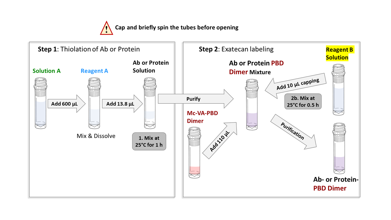
Protocol
Schematic workflow diagram for preparing Antibody- or Protein-PBD dimer conjugates with Val-Ala linker, starting with 20 nmol of antibody or protein (Reagent volume will vary if the amount of antibody or protein is less than 20 nmol).
Frequently Asked Questions:
If you can’t find the answer you’re looking for or need information on general topics, please visit the main Frequently Asked Questions (FAQs) section.
Related Products
Hydrophobic Interaction Chromatography (HIC) Buffer Set (Product#: CM02025): For conjugate prepared via surface amine labeling of the antibody or protein, hydrophobic interaction chromatography (HIC) HPLC can be used to determine whether labeling has occurred. However, due to the highly heterogeneous nature of surface amine labeling, antibodies or proteins with the same DAR or DPR may exhibit slight differences in hydrophobicity. CellMosaic's HIC buffer set can be used with any HIC column. If you do not have access to an HPLC facility, you can send your sample to CellMosaic for analysis.
Size Exclusion Chromatography (SEC) HPLC Protein Standards (Product #: CM92004 and CM92005): SEC HPLC separates the conjugates by apparent molecular weight (MW) or size in aqueous solution. The larger the MW of the conjugate, the earlier it elutes. By comparing the SEC profile of unlabeled IgG and the ADC, you can estimate how much aggregation is in the ADC. CellMosaic® offers two SEC standards for our customers to use with any SEC column. The CM92004 product sheet contains all of the information and methodology you need to run an SEC HPLC analysis. PBD dimer with Val-Ala is highly hydrophobic. This kit is designed to minimize the aggregation and precipitation issues typically encountered with PBD dimer labeling. However, you may still notice some solid precipitate or conjugate aggregation during the reaction. The precipitates will be removed during purification. Depending on the properties of your antibody or protein, recovery may range from 40-80%. If you are concerned about aggregation, you can use size exclusion chromatography (SEC) to assess the extent of aggregation. SEC separates conjugates based on apparent molecular weight (MW) or size in aqueous solution. Larger MW conjugates elute earlier. By comparing the SEC profile of unlabeled antibody or protein to that of the conjugate, you can estimate the level of aggregation in the conjugate. If you do not have access to an HPLC facility, you can send your sample to CellMosaic® for analysis.
ADC stabilizing PBS buffer (5x) (Product #: CM02022): CM02022 contains 5x PBS buffer and other stabilizers to prevent the hydrophobic drugs from interacting with each other during storage, which would cause the ADCs to precipitate out. Stabilization buffer also helps preserve the structure of the ADCs during lyophilization. The buffer is biocompatible and can be used directly for any in vitro and in vivo studies.


 outside of boxes only.")


